Tissue Microarray Technology
Tissue Microarray Technology
Advantages
Tissue microarrays (TMAs) offer significant advantages over conventional histological methods, making them invaluable in medical research and diagnostics. TMAs enable high-throughput analysis by allowing hundreds to thousands of tissue samples to be studied simultaneously on a single slide. This efficiency reduces processing time and costs, making TMAs a resource and cost-effective option.
One of the key benefits of TMAs is the standardization and reproducibility they bring to experiments. By consolidating multiple tissue samples onto a single recipient block, variations arising from different slides or staining protocols are minimized, ensuring consistent and reliable results. TMAs also facilitate comparative studies, enabling direct comparisons between different tissues, disease stages, or subtypes on the same slide, thus aiding in the identification of biomarkers and potential therapeutic targets.
Another advantage of TMAs is their utility in biomarker discovery. Researchers can efficiently screen for potential biomarkers associated with diseases or treatment responses by simultaneously analyzing multiple samples under identical conditions. Additionally, TMAs preserve precious and rare samples, as they require only small amounts of tissue for analysis, making them ideal for studying limited or difficult-to-obtain specimens.
Furthermore, TMAs play a crucial role in validating research findings across larger patient cohorts, enhancing the statistical significance of results. They also serve as quality control tools for immunohistochemistry and in situ hybridization assays, allowing researchers to assess the accuracy and specificity of their staining protocols.
Overall, tissue microarrays offer a streamlined approach to medical research, enabling researchers to make significant advancements in understanding diseases, identifying biomarkers, and developing personalized treatment strategies. The high-throughput, cost efficiency, standardization, and ability to preserve valuable samples make TMAs an essential tool for accelerating research and improving patient outcomes
Applications
Tissue microarrays (TMAs) are widely applied in various fields due to their versatility and efficiency.
Cancer Research: TMAs have revolutionized cancer research by enabling the simultaneous analysis of numerous tumor samples. Researchers can study tumor heterogeneity, tumor grading, and prognosis across various cancer types. TMAs play a crucial role in identifying potential biomarkers associated with specific cancers, aiding in the development of targeted therapies and personalized treatment approaches.
Biomarker Discovery: TMAs are instrumental in the discovery and validation of biomarkers relevant to various diseases, including cancer and other complex conditions. By efficiently screening multiple tissue samples for the expression of specific proteins or RNA molecules, researchers can identify biomarkers that may serve as diagnostic or prognostic indicators, improving patient management and outcomes.
Immunohistochemistry (IHC) Profiling: IHC is a widely used technique for visualizing and quantifying protein expression in tissues. TMAs streamline this process, allowing researchers to screen large cohorts of samples for specific protein markers simultaneously. IHC profiling using TMAs provides valuable insights into protein localization, expression patterns, and cellular interactions, aiding in disease characterization and therapeutic targeting.
Drug Development: TMAs are utilized in preclinical and clinical drug development studies to assess the effects of new drugs on tissue samples from different patient cohorts. By studying the response of tissues to experimental treatments, researchers can gain critical information about drug efficacy and specificity, facilitating the development of novel therapies.
However, these applications represent only a subset of the diverse uses of TMAs in medical research. TMAs also find relevance in fields such as molecular pathology, neuroscience, tissue engineering, and various disease-specific studies. Their high-throughput capabilities and ability to analyze multiple samples simultaneously make TMAs a valuable asset in accelerating research across a broad spectrum of disciplines.
Publications about the technology and discussion of applications and limitations can be found on our Recommended Reading page.
Tissue Arrayers
What is more, Our tissue microarray instruments are designed and developed by Beecher Instruments. Therefore, these instruments allow you to inspect multiple slides that contain hundreds of individual tissues.
Instead of inspecting samples one slide at a time, tissue microarray allows you to work with hundreds at the time. For example, an analysis from 600 different tissues can be completed with just 1 day.

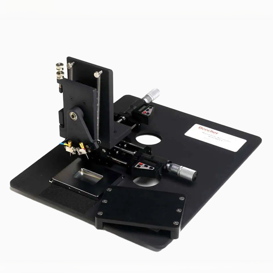
Manual Tissue Arrayer MTA-1
The Manual Tissue Arrayer MTA-1 allows for relatively easy construction of tissue microarrays (TMAs) with several hundred specimens.
The Manual Tissue Arrayer MTA-1 is the entry-level equipment necessary to start up a productive TMA facility. It is an affordable, manually operated high-precision arraying equipment. Skilled users can create arrays with more than 500 samples per slide with this unit.

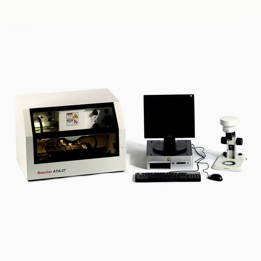
Automated Tissue Arrayer ATA-27
The Automated Tissue Arrayer ATA-27 is designed for fast, accurate and reliable construction of high-density tissue microarray blocks.
The Automated Tissue Arrayer ATA-27 is intended for laboratories requiring higher throughput in array production. It eliminates the tedious manual punching procedure from array construction workflow. After planning and designing the array layout using dedicated software, ATA-27 creates array blocks automatically using robotics to measure block heights, retrieve donor tissue cores, create holes in paraffin matrix and deposit tissue cores into holes.
